Orthopaedic surgeons usually use X-rays and computed tomography (CT) scans to look at bones during the healing process and try to judge when healing is complete. Hannah Dailey’s research team in the Orthopaedic Biomechanics Laboratory is developing methods for using CT scans to objectively measure structural bone healing using virtual mechanical tests. Images from each CT scan are used to build patient-specific models that can be subjected to different types of simulated loads. The results reveal how close the bone is to recovering its structural integrity after injury. This research has applications in the diagnosis of complications such as nonunion (failed bone healing) and to study compromised bone healing, such as in patients who smoke cigarettes.
Below: These patients all suffered shinbone fractures and were CT-scanned at 12 weeks after surgery. Virtual mechanical testing was used to measure how much healing had occurred.
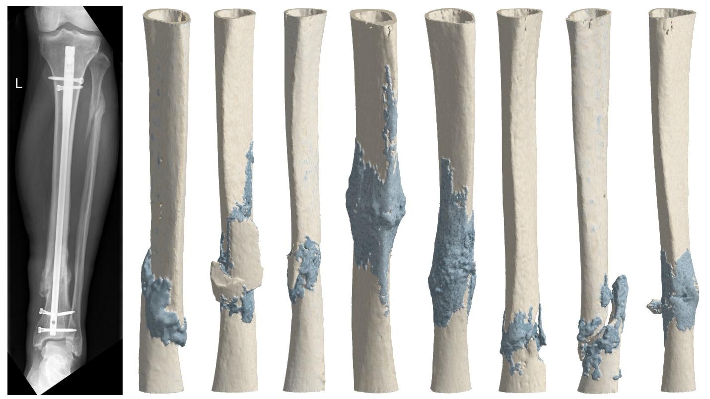
This research was led by Prof. Hannah Dailey in collaboration with investigators at Cork University Hospital (Cork, Ireland) and the Musculoskeletal Research Unit (MSRU) at University of Zurich (Switzerland). Her research to develop virtual mechanical testing methods for measuring bone healing is supported by a National Science Foundation CAREER Award and the Orthopaedic Trauma Association. The work was also enabled by an individual award to lab alum Peter Schwarzenberg, PhD who won an IIE GIRE Fellowship that allowed him to spend six months in Zurich during his PhD studies where he worked on developing a material model for ovine cortical bone.
Read more about Prof. Dailey’s work on virtual mechanical testing in a large animal model and in human tibial fractures.