He had lined up a job at an engineering company, paid a security deposit on an apartment, and was weeks away from making his post-graduation, cross-country move to Seattle when he called his parents.
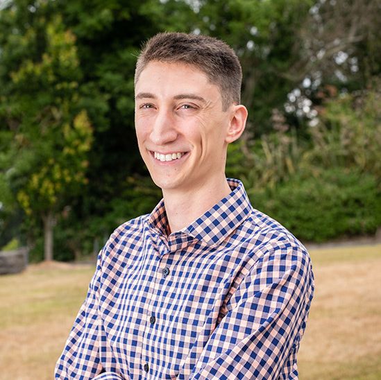
“I was driving to my grandparents on Mother’s Day, and my parents were, too, so it was a good time to talk,” says Peter Schwarzenberg ’16. “I was like, ‘I think I’m going to go to grad school.’ And there was just...silence.”
That was the spring of 2016. Today, Schwarzenberg is a third-year PhD student in mechanical engineering, and one of just 10 students selected from across the country for the Institute of International Education’s Graduate International Research Experiences (IIE-GIRE) program.
As a grantee, Schwarzenberg will spend six months in Switzerland at the University of Zurich’s Musculoskeletal Research Unit (MSRU) developing new mechanical models for callus, the bony tissue that forms during fracture healing.
The IIE-GIRE is a new program funded by the National Science Foundation. Its goal, according to its website, is to “support U.S. engineering graduate students and their academic advisors, for graduate student research experiences abroad.”
Grantees in this first cohort of the program received funding for up to six months to conduct innovative research and promote meaningful connections between their home and host institutions. Stipends are provided for travel, living expenses, health insurance, visa fees, and educational materials. Each student’s advisor also receives $2000 for travel to the host institution or to support similar international endeavors in their home campus graduate programs.
‘A pivotal move’
Schwarzenberg may not have pursued graduate school—and momentarily stunned his parents—if he hadn’t been a chronically hurt runner. He came to Lehigh on a partial scholarship as a track and cross-country athlete.
“In track, my coach encouraged me to try the steeplechase, but I’m from West Virginia, and we didn’t have that in high school,” he says. “All of a sudden an injury-prone kid starts jumping a lot, and I just kept injuring myself.”
After three years, he left the team. It was a pivotal move, he says.
All of a sudden, he had hours free to pursue other interests. He spent the summer between his junior and senior year working with a fellow student on conceiving and building a working prototype for a portable IV device as part of the Baker Institute for Entrepreneurship, Creativity, and Innovation.
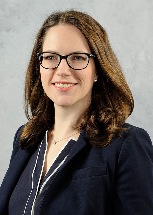
It was during those late nights experimenting with the device that he realized he was doing exactly what he wanted to do—building stuff that could improve people’s lives. During the fall of 2015, the start of his senior year, Schwarzenberg took a class with Hannah Dailey, an assistant professor of mechanical engineering and mechanics. “I was trying to prove something,” he says. “I tried really hard in her class.”
That spring, Dailey offered him a position as a PhD student in her lab.
Counting on callus
As much as he loved building stuff, Schwarzenberg soon found his passion lay in the computational side of research. “I’m definitely a computer nerd,” he says. And for the past 18 months, he’s put his nerd skills to work helping Dailey develop a virtual technique to determine the extent of healing in tibial bone fractures.
Most people who break their tibia, or shin bone, proceed along a normal healing timeline. As the weeks go by, more and more new bone called callus forms along the fracture line. Callus starts out as a spongy material that over time hardens into bone that is just as strong—or stronger—than it was before the break. Patients typically come in for X-rays at regular intervals of, say, three, six, and 12 weeks, and as long as the images reveal there’s increasingly more callus in the region, all is well.
But some people don’t heal normally. This failure to heal is called a nonunion, and it can be utterly debilitating, both from a pain and quality of life perspective. The problem, however, is that surgeons typically rely on X-rays to determine the cause of a nonunion. But X-rays can only reveal so much. They’re two-dimensional. They can be fuzzy. They may provide an incomplete picture if something is blocking the problem area.
It can therefore be difficult for a surgeon to tell the difference between no new bone forming at all—or new bone forming, just very, very slowly. And that difference is important. If no new bone is forming, a second surgery is imperative. But surgery is expensive and carries risk. If new bone is indeed forming, just slowly, it may be better for the patient to wait and avoid the risk of another operation. But a surgeon can’t wait too long, because that can be detrimental, too.
It’s a nuanced problem that requires a new solution. A solution that uses engineering tools to better answer the question of how well a bone is, or is not, healing.
Testing bone like a bridge
Schwarzenberg and Dailey recently published two papers describing how they’ve used virtual structural analysis of low-dose computerized tomography (CT) scans to predict healing in tibial fractures.
CT scans, as opposed to X-rays, provide a highly detailed, three-dimensional picture of what’s going on inside a patient. The scans can also be used to build models.
Using specialized, commercially available software on the CT scans of patients with lower leg breaks, Schwarzenberg built 3-D mechanical structural models that identified the different regions of both the bone and the new bone, or callus. He then ran those models through finite element analysis software—the same program civil engineers use to simulate, for example, how much deformation or movement happens to a structure (like a bridge) when a load is applied to it (like a bunch of cars or pedestrians). Schwarzenberg and Dailey wanted to do the same thing for bones—apply a force and see how much the bone flexed. The less it flexed, the more healed it was.
Schwarzenberg used the finite element software to divide each bone model into tiny zones called tetrahedra that all have a mathematical relationship to each other. He and Dailey then simulated fixing the bottom of the bone so it couldn’t move and putting a load on the top of the bone in the form of a one-degree rotational twist. The technique is called a virtual torsion test.
“So we know what’s happening to the tetrahedra at the edges of the bone,” says Schwarzenberg. “But finite element allows you to calculate what is happening at the neighbors of those tetrahedra and then the neighbors of those tetrahedra, and it calculates all the way through until you’ve calculated every piece inside the bone.”
Those calculations revealed how much the bone flexed when it was twisted.
After brainstorming with Dailey over how to normalize the results to each patient, Schwarzenberg then used the CT scans to digitally re-create a healthy version of each person’s leg. He performed the same virtual torsion test on that healthy model then measured the flex of the unbroken leg against the fractured leg. The resulting percentage helped them determine how stiff the broken bone was compared with the healthy bone. The stiffer a bone was early in the healing process, the quicker the patient could bear weight.
In the end, Schwarzenberg and Dailey found the virtual mechanical test could accurately predict how long it would take a patient to heal. Currently, there are no other tools that can do that. If a patient isn’t healing, it’s an educated guessing game on the surgeon’s part as to why that is, how long the healing process might take—if it takes place at all—and whether to wait or intervene surgically.
The pair’s ultimate goal is to produce a diagnostic test that can help surgeons determine if an additional operation is necessary.
A ‘revolutionary’ approach
“There’s one big flaw in our experimental design,” says Schwarzenberg. “There’s a lot of data in the literature for the mechanical properties of bone. It’s easy to get cadaver bones. It’s next to impossible to get cadaver bones with callus. Bone is this nice, organized, hard structure, and callus is almost like cartilage, kind of soft. It remodels into bone, but at the time points we’re looking at, we don’t expect the callus to have the same underlying structure as bone. We think we’re making it too strong—closer to bone than it should be—because we’re using a model that was developed from only bone. So every time we present somewhere, someone in the audience shoots up their hand, and says, ‘Isn’t callus completely different than bone?’ So our goal is to characterize that callus.”
And that is exactly what he’ll be doing as a 2019 IIE-GIRE fellow, in a country renowned for research into orthopaedic trauma, says his PhD advisor Dailey.
“Switzerland is considered the birthplace of modern surgical fracture fixation,” says Dailey, who is affiliated with Lehigh's Institute for Functional Materials and Devices (I-FMD) and has a long working relationship with researchers at the University of Zurich. “They are world leaders in animal models for bones and how they heal.”
While at the university’s Musculoskeletal Research Unit, Schwarzenberg will be building on research he and Dailey did with the group in 2018. For that project, Schwarzenberg conducted virtual mechanical testing on super-high-resolution CT scans of sheep tibiae, provided by MSRU, to test the effect of a stabilization device on bone healing. As an IIE-GIRE fellow, Schwarzenberg will combine his and Dailey’s virtual technique with an optimization algorithm to refine their understanding of the mechanical properties of callus.
Dailey stresses that this ability to answer questions of whether or not a bone is healing and when it may be capable of bearing weight is revolutionary.
“These advanced modeling and simulation techniques are providing the opportunity to answer fundamental questions like, ‘What are the mechanical properties of newly formed bone?’ Questions that, believe it or not, haven’t been addressed before. Because it’s not like you can take a person, cut out a uniform piece of material, then put it in a machine and test it,” she says. “That’s impossible. But now we can do that in a virtual way.”
Schwarzenberg, now in Switzerland, is proud of landing the fellowship. He’s flattered that he’s one of just 10 recipients. But he is convinced that such recognition hinges on the fact that very high-level research is being conducted every day at Lehigh, and specifically, in Dailey’s lab.
“We’re doing world-class research here,” he says. “We’re working with a world-class animal research institute in Switzerland, and making a difference with what they’re doing. We’re very new, but we’re turning heads at conferences, and we’re butting heads with the big dogs.”
—Christine Fennessy is staff writer for the P.C. Rossin College of Engineering and Applied Science