Scientific progress has provided a solid understanding of the anatomy of the brain. However, there is still no reliable way to examine neuron to neuron communication, as it happens--a key to understanding the correlation between brain structure and brain function.
Chao Zhou, assistant professor of the bioengineering program and the department of electrical and computer engineering at Lehigh University, likens our current brain-mapping ability to a Global Positioning System (GPS) that can help a user locate a city, but cannot offer a street-level view. With current imaging techniques, researchers can observe layers of the brain and understand how different parts relate to each other - but they cannot examine the activity of a single neuron or network of neurons in live tissue. There is no "street-level" map of a living brain.
And yet without such a map, medicine remains in the dark about the most effective ways to treat, prevent, and cure brain disorders like Alzheimer's, schizophrenia, autism, epilepsy, and traumatic brain injury.
President Obama's BRAIN initiative aims to fast-track the search for enlightenment. Announced in 2013, the initiative supports the development and application of innovative technologies that can create a dynamic understanding of brain function with the aim of helping researchers uncover the mysteries of brain disorders.
One very important step in achieving the initiative's goals is the development of optical techniques that are sensitive enough to image at the level of single-neurons and fast enough to image live tissue.
Most neural recording techniques currently used use electrodes or require that neurons be labeled with a genetic or chemical probe in order to view neuronal action potential (the sending of electricity from one neuron to another). These methods have their downsides, according to Yevgeny Berdichevsky, also an assistant professor of electrical and computer engineering at Lehigh and principal investigator in the Neural Engineering Lab.
"While effective, it is not clear that electrodes are able to pick up activity in every neuron--it is also quite invasive," says Berdichevsky. "A marker introduced chemically has the potential to be toxic. It also might be too slow to pick up all the action potentials. Brain imaging cannot currently be done genetically in humans due to safety concerns."
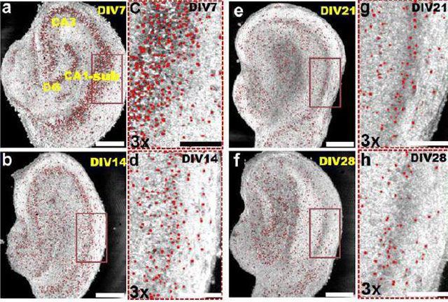
To tackle this 21st century challenge, Zhou and Berdichevsky have joined forces to explore the use of a noninvasive, ultra-high speed biomedical imaging technology, known as space-division multiplexing optical coherence tomography (SDM-OCT). The technique could potentially map the brain using light--and does not require that neurons be labeled.
Originally developed for use in ophthalmology, optical coherence tomography (OCT) works by capturing light as it reflects off the surface of living tissue. According to Zhou, more than 30 million people world-wide per year have their eyes examined using OCT, a non-invasive test that uses light waves to take cross-section pictures of the retina.
Zhou is pioneering a key improvement to OCT that will enable a faster, more sensitive exam to detect eye diseases such as macular degeneration and glaucoma. His patent-approved diagnostic instrument uses a technology called space-division multiplexing to achieve imaging at ten times the speed of current OCT imaging.
The National Institutes of Health have awarded Zhou and Berdichevsky a grant to explore the adaptation of OCT to achieve large-scale imaging of neural activity at the single cell level--a potential game-changer in brain imaging.
Read the full story from Lehigh University on EurekAlert!