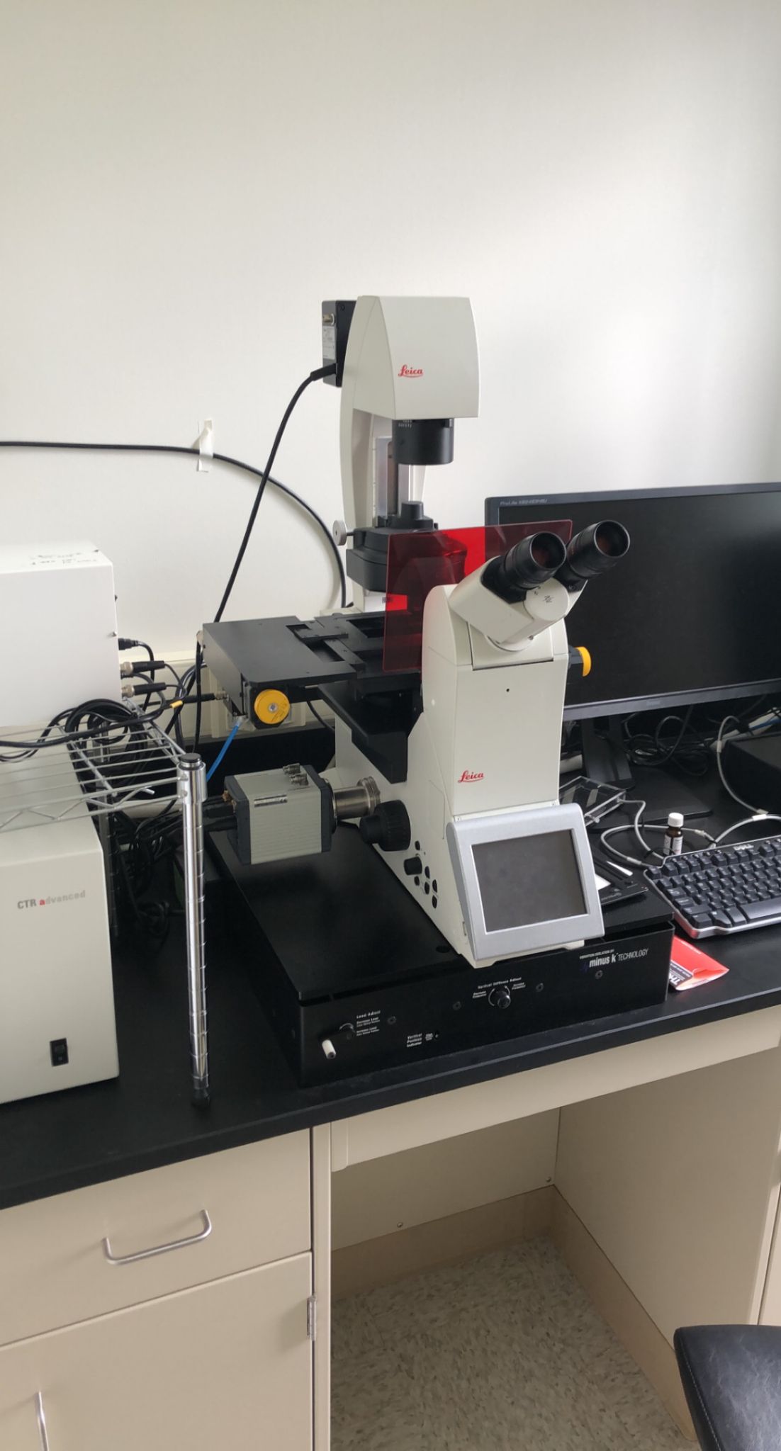
Leica DMi8 Fluorescence Microscope
|
A fully motorized inverted fluorescent microscope. It has remote control operation with a control unit for focus and stage position, LCD touch screen to control easily the microscope light path, objectives, filters, shutters and illumination intensity, and transmitted light LEDs. It can be used for multi-channel fluorescence imaging, imaging of histological samples, Z-sectioning, Z-stack collecting, and time lapse recording. Location: HRH/IAC 354 |
Nikon C2+ Laser Scanning Confocal Microscope with Spectral
|
The C2+ confocal microscope is a basic model within the family of Nikon confocal products. The C2+ is designed as an essential microscopy tool for the laboratory, providing powerful and robust imaging capabilities. The high-efficiency scan heads and detectors, coupled with Nikon's unrivaled optics, provide superior confocal images. The high-speed galvano scanners, operating at rates of up to 100 fps*, enable even the fast-beating motion of cardiac muscles to be captured with precision. The system also provides simultaneous acquisition of three fluorescent channels plus DIC in a single scan. For research that requires spectral imaging capabilities, Nikon's C2si+ system provides dedicated spectral detector units in addition to the standard fluorescence detector units. The C2si+ system allows high-precision and high-speed 32-channnel spectral imaging or high-sensitivity spectral imaging. Built on a reputation of incredible stability and operational simplicity, coupled with superior optical technologies, the C2+ confocal system is the essential laboratory tool. Location: HRH/IAC 348 Fee: $25/h |

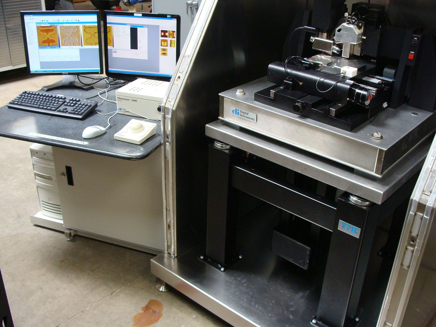
Bruker Dimension V Atomic Force Microscope
|
Location: HRH/IAC 153 Fee: $15/h |
The Dimension family of AFMs has enabled more published data than any other large-sample AFM platform, gaining an iconic reputation in both research and industry in the process. The Icon takes the platform to a new level of excellence, providing higher performance and faster results. The software’s intuitive workflow makes performing even the most advanced AFM techniques much easier than ever before. Icon users achieve immediate high-quality results without the usual hours of expert tweaking. Every facet of the Dimension Icon — from wide-open tip and sample access to preconfigured software settings — has been specifically engineered for trouble-free operation and surprising AFM ease of use. |
Anton Paar SAXSpace Small Angle X-Ray Scattering System
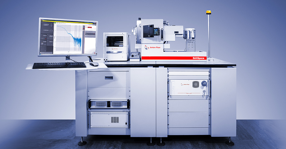 |
A compact lab-scale system for SAXS and WAXS studies with a brilliant block collimation system ensuring an intense X-ray beam for very short measurement times. It determines the size, size distribution, and shape of nano-sized particles and sample domains and is especially suited for analyzing isotropic, colloidal, and biological samples (BioSAXS). SAXSpace’s robust design, high system uptime, and automated workflows ensure a large sample throughput and high-quality SWAXS results. Location: Snyder/IAC 352 |
Customized Optical Coherence Tomography/Microscopy
 |
Optical coherence tomography (OCT) is an imaging technique that uses low-coherence light to capture micrometer-resolution, two- and three-dimensional images from within optical scattering media (e.g., biological tissue). It is used for medical imaging and industrial nondestructive testing (NDT). Optical coherence tomography is based on low-coherence interferometry, typically employing near-infrared light. The use of relatively long wavelength light allows it to penetrate into the scattering medium. Confocal microscopy, another optical technique, typically penetrates less deeply into the sample but with higher resolution. Location: HRH/IAC 350 |
Thermo Fisher LTQ-XL Liquid Chromatograph Mass Spectroscopy
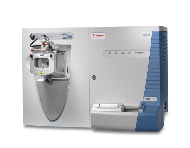 |
The LTQ-XL ion trap MS extends the MSn performance of the LTQ ion trap. It has unparalleled sensitivity and unmatched MSn spectral quality; Pulsed Q dissociation (PQD) for generating more fragments and extending the low mass range; high-resolution isolation (HRI) enables isolation of isobaric mass peaks for cleaner MSn spectra; ultra-fast polarity switching for high sensitivity analysis of unknowns; automated data-dependent neutral loss for PTM and biotransformation analyses; and excellent charge state determinations with Ultra ZoomScan™
It supplies outstanding sensitivity and abundant structural information for protein identification and quantitative differential expression analysis; biomarker studies and PTM identification; metabolite identification and quantification; tissue imaging; and high-speed LC methods. Location: Pashuck/IAC 347 |