Infectious diseases that attack the lung and cause it to fill with fluid present medical science and engineering with a Catch-22 scenario, says Samir Ghadiali.
The fluid causes shortness of breath, wheezing and the sensation of drowning. Anxiety and panic set in. The patient requires immediate medical attention.
The only available remedy is a mechanical ventilator that pushes air into the lungs, breathing for the patient until he can breathe on his own. But the machine further harms air passages and causes a 40 percent mortality rate after several days of use. The phenomenon, ventilatorinduced lung injury (VILI), contributes each year to the deaths of tens of thousands of Americans.
Ghadiali, the Frank Hook Assistant Professor of bioengineering in Lehigh’s department of mechanical engineering and mechanics, believes the optimum treatment for VILI lies in a sound application of engineering principles. He and his students employ computational modeling and lab experiments to gain a clearer picture of the vital functions of the lung and the punishment it endures from VILI.
Ghadiali’s goal is twofold. A better understanding of the biological mechanisms of lung-cell injury during VILI, he says, can lead to the development of drugs that protect lung cells and improve the survival rates of patients undergoing ventilation. Ghadiali also wants to develop a biomimetic drug-screening system that evaluates the impact of air bubbles on cells during ventilation and measures the degree to which that impact is moderated by drug candidates.
“We believe there may be cell-based therapies that can improve patient survival rates,” says Ghadiali. “If you have to undergo ventilation, it may be possible to first take a drug that makes your cells less susceptible to damage during ventilation. Your cells will still be struck by the sledgehammer of passing air bubbles, but they are more resistant to it. This would minimize cell injury. Your lungs can then recover on their own and you can go off the ventilator and survive.”
Life in the deep lung
Ghadiali’s study of pulmonary mechanics, which is funded by a Parker B. Francis Fellowship and a grant from the American Heart Association, focuses on the “deep lung.” There, at the tips of the lung’s elaborately branching airways, micron-sized sacs called alveoli expand and contract when a person breathes, diffusing oxygen into the blood while drawing carbon dioxide from it.
A thin layer of epithelial and endothelial cells, called the alveolar capillary barrier, separates the alveoli from the bloodstream. A severe disease or injury can break down this barrier, allow blood and bacteria to leak into the lungs and prevent the exchange of O2 and CO2. A mechanical ventilator restores that exchange, but at a cost.
“The ventilator saves your life,” says Ghadiali, “but exacerbates the lung injury. We’re trying to understand why the ventilator damages the thin layer of epithelial cells and how that can be prevented.”
Medical researchers have attempted with little success to minimize ventilator forces with surfactant, a compound that helps premature babies breathe.
Going against the grain
Ghadiali has taken the opposite approach. Rather than modify the mechanical forces created by ventilation, his group seeks to modify the responses of cells so they better survive the trauma of ventilation.
To test its hypothesis, Ghadiali’s group accounts for an array of interdependent variables – the functions of lung, blood cells and alveoli, and the mechanical responses and interior activities of cells. This complexity can best be studied, Ghadiali says, by integrating experiments in the laboratory with mathematical modeling on the computer.
“Modeling allows us to study phenomena that are too complex to investigate systematically in the lab. It also enables us to manipulate parameters that are difficult to manipulate in the lab. Models can suggest new lab experiments that might unveil important information.”
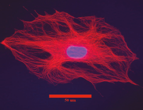
Ghadiali’s students use a software program to simulate the response of alveolar epithelial cells to the shear stress and pressure gradient forces imposed by passing air bubbles. They collaborate with Lehigh biologists to measure the response of cells to these forces, growing and staining a “confluent” group of fully grown cells and a “subconfluent” group of immature, less densely packed cells. They compare the lab data with the modeling results, improve the models and run new tests.
The group’s results have been surprising, says Ghadiali.
“We thought that placing more force on cells would kill larger quantities of cells, but we have found that in some cases that is not true. Like others, we also assumed that a cell modified to become more rigid would better resist certain types of forces. But the truth is more complicated.”
Computational modeling – and what Ghadiali calls a “minimalist approach” to the technique – helps explain some of the counterintuitive results. Ghadiali and his students build their models one level of complexity at a time, choosing only those variables they think are critical to the phenomenon they are examining, rather than attempting to account for every variable at once.
Viscoelasticity is vital
This “piece by piece” approach has helped reveal the lifesaving potential of viscoelasticity for epithelial cells, he says.
“We have found that a cell’s ability to resist force depends not only on how rigid or soft the cell is but also on how viscous it is, that is, how much it behaves like a liquid and how much it moves or deforms over time. In modeling the forces imposed on epithelial cells, we found that only when we added viscosity to the cells did we get results consistent with our lab experiments.”
Ghadiali’s group has confirmed the cells’ viscosity by using “optical tweezers,” a laser tool that oscillates the cell’s cytoskeleton to measure its mechanical properties. The group collaborates with Daniel Ou-Yang, professor of physics at Lehigh and a pioneer in developing the tweezers.
“We’ve found that in addition to making cells softer, some treatments can also make cells viscous,” says Ghadiali. “This viscoelasticity enables a cell to ‘damp out’ the transient forces imposed by microbubble flows in the deep lung.
“The fact that we can manipulate this viscoelasticity suggests to us that it may be possible to develop pharmaceutical compounds that alter the viscoelasticity of the cells so they better withstand the forces of VILI.”
Ghadiali’s group is building a biomimetic, microfluidic system that contains flexible, branching air passages and models the entire alveolar-capillary barrier. The goal, he says, is to develop an in vitro system that prescreens drug candidates before they undergo in vivo testing.