Ever since July 20, 1969, when Neil Armstrong left the first human footprint on the surface of the moon, scientists have been fascinated by the fine powdery soil in which that impression was made. Today, more than 40 years later, Carol and Christopher Kiely are using a new imaging technique called X-ray ultramicroscopy (XuM) to examine the internal structure of the lunar soil particles that were collected from the Sea of Tranquility during the Apollo 11 mission.
“These particles are like tiny time capsules,” says Carol Kiely, an adjunct professor in the department of materials science and engineering. “They provide us with clues to all the geological processes that have occurred on the lunar surface for the past 3.5 billion years.”
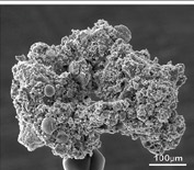
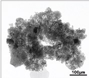
To the naked eye, lunar soil is a fine charcoal gray powder. Under a microscope it becomes a collection of tiny rock, mineral and glass fragments of all shapes and sizes. This assortment, Kiely says, owes its origin to the fact that the moon, unlike the earth, has no atmosphere to protect it from solar wind or from micrometeors that smash into its surface at velocities of up to 25 km per second. This continuous bombardment, over billions of years, has led to the formation of lunar soil, or regolith.
The huge amount of energy behind a micrometeor impact, says Kiely, can fracture the underlying rock and cause localized melting. Splashes of molten regolith can then form droplets, which cool and solidify to form glassy spheres, ellipsoids, teardrops and dumbbells before returning to the lunar surface.
Some of this molten regolith returns to the surface before cooling and seeps down in between the underlying soil. As it cools, it encases the tiny mineral and other fragments in a glassy matrix, forming a type of particle called an agglutinate that is not found anywhere on Earth. The rise in temperature also releases trapped gases implanted by the solar wind, causing bubbles to form in the molten regolith. As a result, much of the glass found on the lunar surface is vesicular.
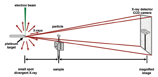
While 40 years of research has revealed much about the physical and chemical properties of lunar regolith, it has been impossible to view the internal structure of a particle without fracturing it or slicing it open. The X-ray ultramicroscope now enables scientists to do this. Originally developed at the Commonwealth Scientific and Industrial Research Organization (CSIRO) in Australia, XuM utilizes the divergent beams of X-rays generated in a scanning electron microscope (SEM) when the electron beam is focused onto a piece of platinum. These X-rays then pass through a lunar dust particle and onto an X-ray detector. Unlike many other microscopy techniques, this method of imaging does not require any focusing optics – the entire XuM image is always in focus – and the resolution, which can be better than 300 nanometers, depends on the size of the X-ray source. This means that for the first time, the entire internal structure of a lunar particle can be imaged with the particle still intact.
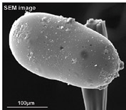
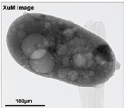
“Combining these new X-ray images with the corresponding secondary electron micrographs has allowed us, for the first time, to view the internal and external structure of the same particle,” says Christopher Kiely, a professor of materials science and engineering and director of Lehigh’s Nanocharacterization Laboratory. “This gives us a much fuller understanding of a particle’s morphology. For example, glassy particles that often appeared smooth on the outside were found to contain a myriad of pores, far more than we expected.”
The XuM’s ability to take a sequential series of images while rotating the sample through 360 degrees yields a more global view of the internal structure of a particle and can help prevent erroneous conclusions being drawn from a single flat 2-D projection image. For example, a platelike inclusion could be mistakenly identified as a needlelike feature when viewed ‘edge-on’ in a single 2-D projection image. Sequential imaging also enables rotational movies to be made, which provides a fascinating 3-D view of a particle’s internal structure.
As part of Apollo 11’s 40th-anniversary celebrations in July 2009, a collection of XuM and SEM micrographs, together with SEM stereoimages, was displayed in the Smithsonian Air and Space Museum in Washington, D.C. Carol Kiely presented the latest XuM/SEM results at the 41st Lunar and Planetary Science Conference in March.