Cervical cancer was once one of the most common causes of cancer death for American women. But death rates fell by 74 percent from 1955 to 1992 and, says the American Cancer Society, continue to drop about 4 percent a year, chiefly because of increased use of the Pap smear.
Nonetheless, it is estimated that over 4,000 American women will die of cervical cancer this year. And the disease is one of the leading causes of death in middle-aged women in the developing world.
Now, a new computer-assisted visual interactive recognition system developed by a research team led by Xiaolei Huang may lead to a more cost-effective method of detecting cervical cancer in its early stages.
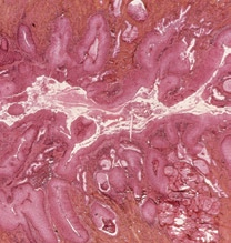
A complementary test to the Pap smear is the cervigram, a digital photo of the cervix. To highlight abnormal tissue, a weak solution of acetic acid is applied. Upon contact with the acid, all forms of precancerous tissue exhibit some degree of opacity, or aceto-whiteness. Various patterns signaling cervical lesion, such as vasculature, mosaicism and punctations, can appear inside the aceto-whitened region. The whitening starts to wear off after about five minutes.
A cervigram enables a medical professional to identify areas containing aceto-whitened tissue. This negates having to send out samples for analysis, which is important when access to wet lab facilities is limited or nonexistent. A cervigram can also be transmitted via the Internet to a professional at a remote site for interpretation.
“Recognizing aceto-whitened areas and other abnormal visual patterns, however, is not as easy as you might think,” says Huang, the P.C. Rossin Assistant Professor of computer science and engineering. “Every patient is different, and the conditions under which images are taken may vary. Our aim is to develop a software system that will facilitate the recognition process and reduce the number of false-positive and false-negative results.”
Such a system, says Huang, must be robust. A simplistic approach might identify pixels of a certain color or intensity and highlight these as affected areas. This would be error-prone as pixels in unrelated parts of the image might have similar characteristics due to glare or other imaging anomalies. Color and contrast ranges will not be the same in every image. “In order to develop a reliable cervigram analysis system,” says Huang, “the texture, size and relative geometry of regions must also be considered.”
Huang’s system combines data derived from images annotated and analyzed by trained medical professionals with computer learning software to reduce false-positive readings. The reliability of the system depends on the number of annotated images used. To help Huang achieve her goal, the National Library of Medicine (NLM) and the National Cancer Institute have given her team access to more than 100,000 anonymous cervigrams and their corresponding diagnostic notes.
Although she is only in the second year of her project, Huang’s software can already reliably identify aceto-whitened areas. Another goal of the project is to enable users to retrieve similar cervigrams, along with diagnostic comments from the NLM archive, via a Web connection. This may help medical personnel who must grade cervical lesions and decide what treatment to take.
“We hope to be able to extend this methodology to other fields of imaging diagnostics such as the analysis of mammograms,” says Huang.
Huang is working with Profs. Daniel Lopresti and Gang Tan in Lehigh’s department of computer science and engineering; George Nagy of Rensselaer Polytechnic Institute; and Dr. Joseph Patruno of the department of obstetrics and gynecology at Lehigh Valley Hospital in Allentown. The work is funded by NSF