For the past 40 years leading experts in the field of electron microscopy and microanalysis have gathered at Lehigh each June to teach students from industry, government labs and universities how to get the best possible data from their own electron microscopes. Their goal is to instill a passion for the subject and pass on their collective expertise to the next generation of microscopists.
To date, more than 5,000 students from 50 states and 36 countries have taken courses at the school.
The idea for the Microscopy School was conceived by Joseph I. Goldstein, then a professor of materials science and engineering at Lehigh. “We had no scanning electron microscope here at Lehigh then,” Goldstein, now dean emeritus of engineering at the University of Massachusetts, recalls. “To convince the university it needed one, I recommended offering a course in microscopy and bringing in an instrument from a company.”
The first school was held in the summer of 1970 with three lecturers, a dozen students and a single scanning electron microscope (SEM). Since then it has grown to become the largest school of its kind.
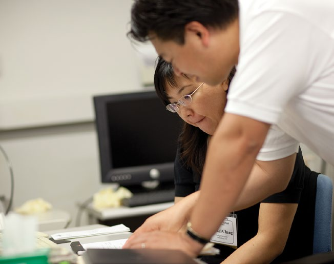
Today, the two-week school offers basic and advanced courses that combine theory with lab practice. Manufacturers’ microscopes supplement the 11 scanning and transmission electron microscopes in Lehigh’s Nanocharacterization Laboratory, one of the largest of its kind in the U.S. This year’s school was attended by 130 students and included contributions from 37 lecturers (12 from Lehigh) and 22 instrument manufacturers.
“These microscopes are extraordinarily powerful platforms,” says lecturer Dale Newbury ’69, a Microscopy Society of America Fellow with the Surface and Microanalysis Science Division of the National Institute of Standards and Technology. “How to interpret the images and how to apply that information usefully – that’s what we’re providing.”
Perhaps the defining mark of the school is the lecturers’ ability to stay current with the latest advances in the field.
“The Microscopy School began only five years after the first commercial SEMs were available,” says Charles Lyman, a professor of materials science and engineering at Lehigh who has taught courses for 35 years at the school. “But it has followed the advances in scanning electron microscopy and microanalysis.”
The newer SEMs can feature advanced X-ray spectrometry; variable-pressure, low-voltage and high-resolution microscopy; electron backscatter diffraction; and focused ion-beam technology. The latest transmission electron microscopes (TEMs) have aberration correctors that improve image and chemical mapping resolution, and electron energy-loss spectroscopy for compositional analysis of lighter elements.
“For a long time the electron microscope was simply an imaging device,” says Newbury. “Now we use it to study morphology, composition and crystallography.”
All of these changes are reflected in the school’s continually updated course work. The school also uses the Internet to remotely access microscopes at other universities.
“A person who took the course some years ago,” says Lyman, “would hardly recognize it now, so much has changed.”
Engineers and scientists of all types attend the school. “Many work in large-scale industrial laboratories that support the mainstream semiconductor and chemical industries,” says Christopher Kiely, director of the Microscopy School and of Lehigh’s Nanocharacterization Laboratory. “Others are from less conventional fields, including forensic scientists from the FBI investigating crime scene evidence and museum conservators seeking to determine the authenticity of paintings.”
Robert Edahl of the NASA Langley Research Center has taken five courses at the Microscopy School. “The faculty,” says Edahl, “are incomparable. They know the latest in their field. Things have changed just in the six years I’ve been attending the school.”
Several definitive textbooks have arisen out of the courses taught in the Microscopy School. For example, Goldstein, Lyman, Newbury and four other lecturers have coauthored Scanning Electron Microscopy and X-ray Microanalysis: A Text for Biologists, Materials Scientists and Geologists, whose fourth edition will be published next year.
“To maintain our position as the world’s premier microscopy school in this ever more competitive arena,” says Kiely, “Lehigh will need to continue to invest in first-class microscopy personnel and state-of-the-art instrumentation.”