Xiaolei Huang, the P.C. Rossin Assistant Professor of computer science and engineering, looks for three things in a research project.
“When I choose a project,” says Huang, “I seek a difficult problem that allows me to push the state of the art in computer science and that also has societal impact.”
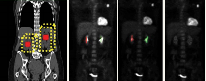
Huang harnesses the power of the computer to process and interpret biomedical images. One of her software programs constructs 4-D computer models – with time as the fourth dimension – that measure the changes in the heart’s structure as it expands and contracts. The models help cardiologists discern the differences between the normal deformations that occur in healthy hearts and the abnormal deformations that occur in damaged and diseased hearts.
In another project, recently funded for a fourth year by the National Library of Medicine, Huang uses object segmentation techniques to index NLM’s database of 100,000 images of cervical lesions. By categorizing lesions according to color, texture, size and shape, she hopes to make it easier for medical personnel to retrieve and study images, and to determine the seriousness of new lesions by correlating more quickly and accurately between image features and diseases. A related project involving the analysis of cervigrams is funded by NSF.
Huang also applies image analysis techniques to help study the cytoskeleton and its role in cell division. The cytoskeleton protects the cell, maintains its shape, enables it to move, and facilitates the intracellular transport of materials. It also governs cytokinesis, the final step in mitosis, during which tiny filaments assemble into a ring, constrict and cleave the cell into two identical daughter cells.
Huang writes algorithms to show how the cytoskeletal filaments form from the protein actin, and interact and interconnect as they assemble. She and her students collaborate with Dimitrios Vavylonis, an assistant professor of physics in Lehigh’s College of Arts and Sciences, who models the physical properties of cytoskeleton and other adaptive biological materials. The project is supported by the National Institutes of Health.
The researchers’ goal is to determine the topology, or physical arrangement, of the filament network and how it changes over time. Their results could be useful in the drive to develop drugs that treat cancer by preventing the division of cancerous cells.
The task is complicated by the ever-changing topology of the cytoskeleton. And the microscopic images of actin filaments have a low signal-to-noise ratio, making it difficult to locate the filaments’ centerline and determine their shape and position in the topology.
“The filaments in the cytoskeleton assemble into a network before they form a ring,” says Huang. “The filaments cross each other, grow and touch each other, and then disassemble. This is an extremely dynamic network.”
Huang and Vavylonis have developed an interactive, open-source software tool called JFilament that visualizes individual filaments, segments them (extracting their centerline and representing their geometry with curves) and tracks them (measuring motion and deformation over time) in 2-D and 3-D. The software is based on a Stretching Open Active Contours algorithm developed by Huang’s Ph.D. student Hongsheng Li.
Active contours are deformable parametric curves that adapt to fit image features. Li uses open curves to represent filaments and has proposed strategies to enable the contours to grow over faint elements and handle crossed filaments. Other methods that segment linear structures produce binary pixel-wise segmentation results and have difficulties reconstructing a curve. In addition to segmenting and tracking filaments and cell boundaries, says Huang, JFilament can utilize the active contours’ results to quantify the static and dynamic properties of cytoskeletal filaments, such as bending and elongation.
The researchers validated the performance of the new software by using it to measure the known properties of simulated filaments. They then used JFilament to measure the dynamic properties of actin filaments imaged with total internal reflection fluorescence microscopy and, for the first time, of actin cables in yeast imaged with onfocal microscopy.
They described their results in a paper that will be published later this year in the journal Cytoskeleton.