Changes in the mechanical properties of biological cells, many scientists believe, are a major contributing factor to the development of bone, kidney and vascular disease.
Measuring the physical properties of individual living cells, however, is not an easy task because of their small size and because measurements must be made in a suitable aqueous medium in order to keep the cells alive.
Direct contact methods for measuring the stiffness of individual cells have involved the use of a cantilever in an atomic force microscope. Noncontact techniques involve magnetic and laser tweezers. Both have achieved success, but they require sophisticated and expensive equipment, and time, to collect necessary data.
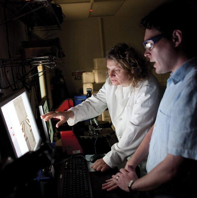
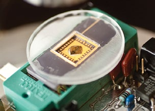
Svetlana Tatic-Lucic, Susan Perry, Sudhakar Neti and Arkady Voloshin are seeking a more rapid and cost-effective method. They have designed a novel MEMS (microelectromechanical system) device to measure the compliance, or stiffness, of a single cell.
“The advantage that MEMS technology has over other systems is that it can integrate both micro-sensors and micro-actuators on a single chip,” says TaticLucic, the principal investigator on the project.
The researchers’ first-generation device incorporates a cell-positioning system, a polymer V-shaped electrothermal actuator (ETA) array capable of compressing a living cell by a desired amount, a sensor to measure the force exerted on the cell, and a thermal sensor.
“We chose an epoxy-based polymer for the ETA primarily because of its large thermal expansion coefficient,” says TaticLucic, an associate professor of electrical and computer engineering who is affiliated with Lehigh’s Bioengineering Program. “Comparing it to an ETA made from polycrystalline silicon, there should be a five-fold increase in thermal efficiency. In addition, the polymer ETA would be easier to fabricate.”
The integrity of any MEMS device is highly dependent on the way it is made. This requires a great deal of skill and attention to detail. Care must be taken to avoid any contamination, no matter how small. The fabrication was carried out in the clean room of Lehigh’s Sherman-Fairchild Laboratory for Solid-State Studies.
“Our initial goal was to find out if the polymer ETA could actually deliver the required compressive strain to a single cell,” says Tatic-Lucic.
To do this, the researchers immersed the entire MEMS device into a liquid medium. Mouse fibroblast cells were then introduced into the cell medium using a micropipette. One of the cells was then trapped in place by applying an AC voltage to the electrodes of the cell-positioning system, a technique known as dielectrophoretic positioning. The cell was compressed by applying another AC voltage to the actuator.
“We found that the cell could be compressed repeatedly and it did not disintegrate during or after the compression test,” says Tatic-Lucic. “The implemented displacement was 4 microns.”
In future experiments, the researchers will use a force sensor to measure the reaction force exerted by the cell during a compression test. This will allow them to calculate the compliance of the cell. The project is being funded in part by the National Aeronautics and Space Administration (NASA).


